Though the clinical diagnosis of gynecomastia is straightforward, there is some issues that may make this seemingly simple issue a little tricky. These diagnoses that mimic a certain other disorder are called differential diagnoses.

One such common differential diagnosis is pseudo gynecomastia. As the name suggests it refers to ‘false male breast’ meaning fat that masquerades as breast tissue. It is especially common in men who are overweight or obese. However in my opinion the term pseudo gynecomastia is overused and over-diagnosed. There have been instances in mine and my colleagues’ practices where a patient is referred to us as pseudo gynecomastia and the patient is expecting liposuction as the sole treatment.
Some surgeons have even gone ahead and done liposuction to this apparently pseudo gynecomastia only to them realize that there is in fact a large amount of breast tissue left. Now, this complicates the issue manifold. One, the patient has not been told that he requires a gland excisions and a bigger skin cut to do the same. Now, either the surgeon has to explain to the patient’s friends or relatives who have accompanied the patient that the surgical plan has changed as he has now noticed breast tissue or he has to end the procedure there and plan breast tissue removal later on. In either of these scenarios, the surgeon does not come off with flying colors. More importantly, the patient is put through a procedure that he is ill-prepared for.
If the surgeon takes the relatives into confidence and goes ahead and operates and removes the breast tissue, it is left to him to explain it to the patient when he comes out of anesthesia. The matter is easier said than done as it depends entirely on how the patient takes the news. If the surgeon chooses the second option and ends the surgery without removing the gland, he needs to explain that to the patient who may not be happy hearing that he needs another procedure to sort out his chest. I have seen patients who underwent only liposuction for ‘pseudo gynecomastia’ and coming to me complaining that their surgeon has left a lot of hard fat under the nipple. I have had to explain such patients that only fat was removed and the gland was left behind and indeed the diagnosis may be wrong. Now imagine the patient’s conundrum at this phase. An issue that could have been easily sorted in the first procedure has now dragged on not only needing another procedure but also making the patient invest a disproportionate amount of time to sort out the one issue at hand.
This is especially true if the patient following liposuction gets fitter and loses more weight only to notice his breast tissue getting more prominent. Before the surgery and his weight loss, the overall smooth contour due to fat might have camouflaged the breast tissue better than when he gets the liposuction or loses weight. All these issues are readily avoided if one is carefully diagnosing pseudogynecomastia. If the diagnosis is correct then liposuction should suffice.
Apart from fat, there may be other swellings on the chest that simulate a gynecomastia. These swellings may arise from any of the structures over the chest. In most of cases they are unilateral, meaning they are usually only on one side. Nonetheless, it is paramount to diagnoses the swelling before proceeding with the treatment plan.
Skin swellings are quite common on the chest too. The commonest of them is the lipoma. It is an accumulation of immature, abnormal fat cells under the skin. They are more mobile under the skin than the breast tissue and are softer. The treatment for these lipomas is excision (removal) through a small cut in the skin. A big enough lipoma can mimic a breast. Many other such swellings can occur on the chest skin like a sebaceous cyst, dermoid cysts, fat necrosis after injury ( a situation where the fat cells die in a particular area following a high impact injury and clumps up) etc.
Most of them are quite straight forward for the doctor to rule them out. But another sometimes serious and under-diagnosed issue is mastitis. It is basically an infection of the breast tissue. It is way more commoner in females but does occasionally affect men with breasts. The breast glands can get infected by various paths. It can get infected through external sources across the skin like an injury to the skin, nipple piercings, or just a skin infection that spreads inwards. The other mode is infection via blood. Any infection in a part of the body can theoretically spread to another tissue like the breast. When one has mastitis they would complain of severe pain, redness over the chest skin, sometimes pus discharge from the nipple, enlarged lymph nodes in the armpits ( axilla), and fever. The treatment is a course of antibiotics and occasionally removal of the pus and if necessary, the gland.
The word ‘Tumor’ incites various feelings in people. They vary from formidable, dread, distressing, and downright scary. But, not all tutors are not cancers and not all cancers are untreatable. Most of the tumors are benign, meaning, non-cancerous. They are treated like any swellings and usually require removal. In the breast tissue, some men do develop breast cysts and fibroadenomas.
Breast cysts are fluid-filled swelling that has a thin capsule. These cyst again are commoner in females and show up a lump on examination. In most cases, ultrasound is required to correctly diagnose. Sometimes your doctor will insert a thin needle into the breast lump and attempts to withdraw (aspirate) fluid. This may be done under ultrasound guidance or by feeling the lump itself. If the fluid comes out and the breast lump goes away, your doctor can make a breast cyst diagnosis immediately. If the fluid is not bloody and the breast lump disappears, you need no further testing or treatment unless it comes back. If the fluid appears bloody or the breast lump doesn’t disappear, your doctor may send a sample of the fluid for lab testing and refer you to a radiologist for the scans. If no fluid is withdrawn, your doctor will likely recommend a scan such as a diagnostic mammogram and or ultrasound. Lack of fluid or a breast lump that doesn’t disappear after aspiration suggests that the breast lump or at least a portion of it is solid, and a sample of these cells may be collected to check for cancer (fine-needle aspiration biopsy). The treatment of breast cysts is rarely by surgery. It is operated upon only if the diagnosis is unclear even with the scans or if it is troublesome to the patient.
Other breast swellings that may occur in a male breast are duct ectasia, fibroadenoma, and breast cancers. While duct ectasia and fibroadenomas are benign, cancer is another matter altogether. In duct ectasia, the patient may have a scary sign of bloody nipple discharge. One fine day he wakes up to find a bloodstain on his nipple. Many men freak out and rush to the doctor fearing cancer. It usually is a duct ectasia which is nothing but an alteration in the ductal structure. It sometimes needs treatment but always needs assessment. Cancer should be ruled out when there is nipple discharge.
Fibroadenomas are the commonest swellings in a female breast. Some do occur in a man. They feel like a firm lump that is unnaturally mobile. It was often referred to as the ‘mouse in the breast’. While that scenario is far fetched for a male breast, nonetheless it depicts how mobile a fibroadenoma can be. The treatment is removal if the size is more than 3cms in diameter. Occasionally men do wait for them to get very big assuming it’s just a skin lump. While it’s not necessarily dangerous, it’s better to wait to know what it is than without.
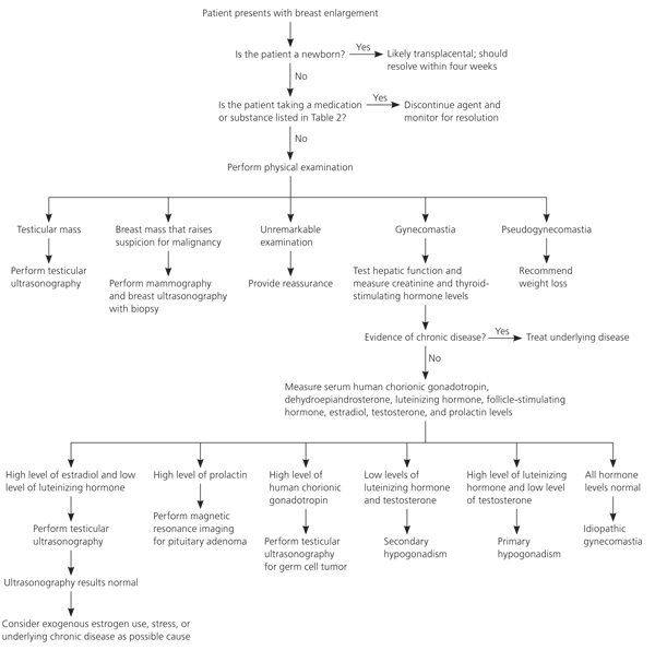
Gretchen Dickson in 2012 published a widely referenced article in the journal, American Family Physician that outlined an algorithm to diagnose gynecomastia.

When one talks about tests for a gynecomastia patient, it has two facets to it
- Tests to check for causes of gynecomastia
- Test mandatory to check if the patient is fit for surgery
Once the doctor suspects its gynecomastia, then the next step is to check if there is indeed a hormonal issue or its idiopathic in nature. The word idiopathic itself means that the cause is unknown. It accounts for over 70-90% of all cases of gynceomastia. However, before diagnosing someone as ‘Idiopathic gynecomastia’ the doctor needs to rule out a few things. The other category of patients belongs to ‘Secondary gynecomastia’ wherein there is a proven cause for gynecomastia as was enlisted above.
Apart from the drug induced gynecomastia that may be easier to diagnoses, the doctor should bear in mind other hormone disorders. Laboratory evaluation is indicated only if the clinical assessment suggests a secondary underlying cause. It is not needed for boys at puberty for enlargement due to fat (pseudogynecomastia) and for men taking drugs known to cause gynecomastia.
In cases of secondary gynecomastia without a clear cause, laboratory tests should be advised and must include, liver, kidney, and thyroid function tests (to exclude the respective causative medical conditions), as well as hormonal tests. The hormonal analysis is always in a series of tests as a part of the evaluation. They include:
- Estrogen level
- Total and free testosterone
- Luteinizing Hormone
- Follicle-stimulating hormone
- Prolactin
- And occasionally hCG, DHEASO4 or 17 ketosteroids, SHBG and αFetoprotein
- If the patient’s testes are small, the patient’s karyotype (chromosomal analysis) should be done to exclude Klinefelter’s Syndrome.
If all tests are negative, the patient should be diagnosed with idiopathic gynecomastia. Sometimes it is indeed advisable to have an endocrinologist look at such patients, as there may be other important than just gynecomastia. It is especially more prudent to meet one if these tests reveal significant variations. Early stages of secondary gynecomastia can be medically treated through the gland does not always regress and may end up needing procedure to sort the gland.
Ultrasonography and mammography can occasionally be used to differentiate fat from breast tissue or if there are abnormal masses especially in terms of consistency. Scans are definitely when the patient has one of the signs suggestive of a cancer lump. Other than these, scans may be necessary also to ascertain breast tissue that feels abnormal or feels irregular in places. Mammography is about 90% sensitive and 90% specific for cancers compared with benign masses in men.
However, a biopsy is the only way to make a definitive diagnosis. Patients with a hard, irregular, or asymmetrical mass, nipple discharge (bloody or non-bloody), enlarged armpit lymph nodes, or a mass fixed to skin or the chest wall must have a biopsy. Usually, a core biopsy is recommended over a fine needle or excisional biopsy. In core biopsy, a small amount of tissue is taken via a small hole under local anesthesia. It is more accurate over the commonly done fine needle biopsy. In a fine needle biopsy, an injection is given and the feels obtained through it are examined under a microscope. Since the amount of cell sone gets from such biopsy is very small, the chances of error are consequently high. An excision biopsy is one where the lump is removed in its entirety and sent for testing. While this is the most accurate, it is reserved for specific cases where the diagnosis with routine biopsies are not confirmatory.
Rarely done tests include:
Ultrasound of testes: If there is any abnormality in the testes on examination, or if there is a raised beta-hCG or alpha-fetoprotein.
Abdominal Scans: If a tumor of the adrenal glands or the testes is thought to be responsible for the gynecomastia after hormonal analysis.
Chest X-ray: If a lung tumor is suspected as a cause for gynecomastia. Sometimes tutors in other tissues may be hormonally active, meaning they may produce hormones which in turn may lead to gynecomastia. Though these are rare, one does come across such cases and a quite often a diagnostic dilemma.
Once these diagnostic tests are done as necessary, the next set of tests are for those planning to undergo the corrective procedures. Since the procedures can be theoretically done under local or general anesthesia, the test also varies depending on the anaesthesia planned. For a gynecomastia surgery under local anesthesia, no additional tests are needed, if the patient is clinically fit and has a clear unremarkable history. When the procedure is planned under general anesthesia a set of tests are mandatory.
- Complete blood counts
- Blood sugar levels
- Coagulation parameters: PT, INR and aPTT
- Kidney function tests: Serum Creatinine and Urea levels
- Other tests that are often done are: HIV screening, HBsAg to check for hepatitis B, HCV etc
- Apart from these routine tests, a few more may be advised depending on the patient’s age, medical history and the factors. These include Echocardiography, Chest X-rays, ECG, Liver function tests, Sickle cell tests etc. Cardiac (heart) evaluation becomes necessary when an older patient comes for corrective surgery. Occasionally a consultation with a cardiologist or the relevant physician is needed before surgery.
This post is an excerpt from Dr Sreekar Harinatha’s highly acclaimed book on gynecomastia titled ‘The Male Breast: What You Should Know about Gynecomastia’. It’s available on Amazon on the link here.
To Book Appointments with Dr. Sreekar Harinatha, call 7022543542…


 WhatsApp us